
Contents
- 1 Overview of Darkfield microscopes
- 2 How does dark field differ from conventional microscopy?
- 3 How Does Darkfield Microscopy Work?
- 4 What Are the Advantages of Using Darkfield Microscopy?
- 5 What Are the Challenges of Using Darkfield Microscopy?
- 6 What Components Are Necessary for Darkfield Microscopy?
- 7 What does my scope need to do dark field?
- 8 What Is Darkfield Microscopy?
- 9 When is dark field good to use?
- 10 Why Is Darkfield Microscopy a Good Imaging Technique?
- 11 History of Darkfield microscopes
Overview of Darkfield microscopes
- Darkfield microscopes are used in metallurgy and gemology to look for imperfections and otherwise hard-to-spot elements that would be otherwise impossible to spot with more traditional bright field microscopes.
- In a darkfield microscope, if you were to look at the back of the objective through a Bertrand lens or phase telescope, it would appear filled with light.
- A darkfield microscope is a brightfield microscope that has a small but significant modification to the condenser.
- Darkfield microscope illuminates a sample only from the sides so it appears bright against a dark background.
How does dark field differ from conventional microscopy?
In conventional bright field illumination, your specimen is lit from a central light source (you can read more about bright field microscopy in this Bitesize Bio article). This results in a large contrast image. However, in dark field microscopy this light source is blocked by a condenser or a ‘stop’ below the stage. This condenser or stop scatters the light allowing only oblique rays to reflect and refract off your specimen which in turn creates a bright image on a dark background.
How Does Darkfield Microscopy Work?
Darkfield illumination requires blocking most of the light that ordinarily passes through and around the specimen, allowing only oblique rays to interact with the specimen.
What Are the Advantages of Using Darkfield Microscopy?
Darkfield microscopy has many advantages. Its dark background offers a high degree of contrast, making it easy to see samples on difficult backgrounds. This technique is easily accessible since many brightfield lab microscopes can be configured for darkfield illumination. Besides the microscopy benefits, darkfield images can even look like works of art with the sample beautifully illuminated.
What Are the Challenges of Using Darkfield Microscopy?
Care must be taken when preparing specimens for darkfield microscopy because the features that lie above and below the plane of focus can scatter light and contribute to image degradation. The cleanliness of the slide is an important factor when imaging, but it is even more important in darkfield since every piece of debris will be illuminated and can take away from what you’re trying to see.
What Components Are Necessary for Darkfield Microscopy?
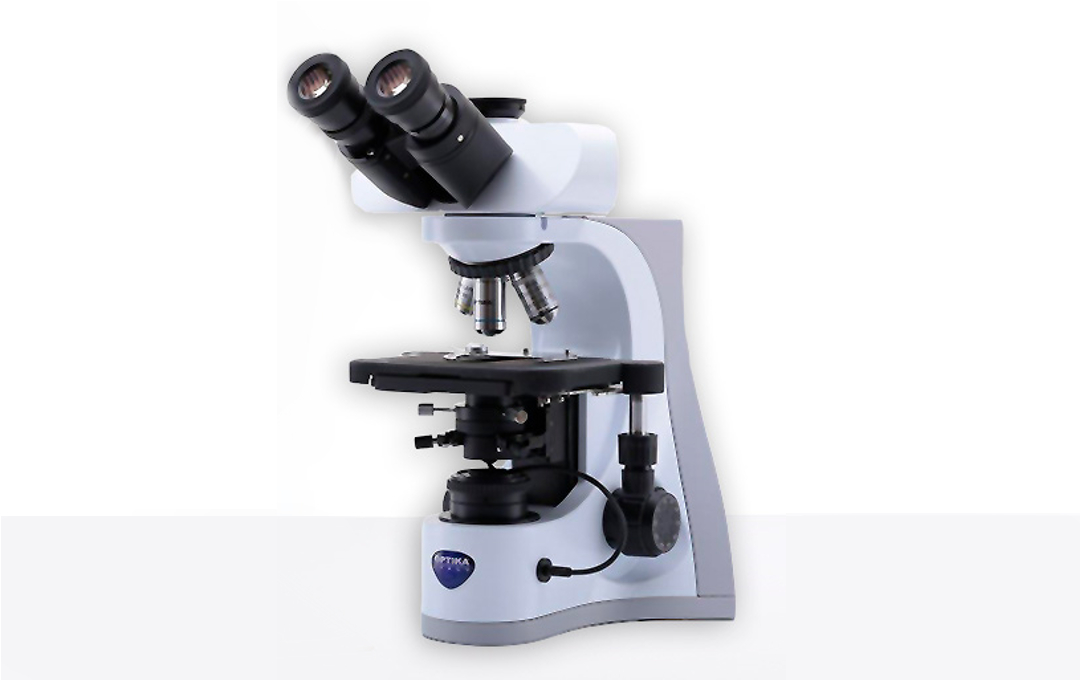
Almost any brightfield laboratory microscope can be easily converted to perform darkfield illumination. The easiest way to do this is to switch out your current condenser with one that is designed for darkfield illumination (Figure 1).
What does my scope need to do dark field?
Most stereo and compound microscopes can do dark field imaging. Check your microscope’s specifications to see if this is your case. If your microscope does not have a built-in condenser or stop, don’t worry, you can probably still use your microscope for dark field imaging. You should be able to purchase an aftermarket condenser or even make your own stop. Read below to learn more about condensers and stops.
What Is Darkfield Microscopy?
Darkfield microscopy is a technique that takes advantage of oblique illumination to enhance contrast in specimens that are not imaged well under normal illumination conditions.
When is dark field good to use?
Dark field is useful when you would like to view unstained, transparent specimens. The best specimens for dark field should have a refractive index that is close to the surroundings and otherwise difficult to image using conventional bright field microscopy. For example, many small aquatic organisms have refractive indices that are very similar to their surrounding water, making them ideal candidates for dark field microscopy. Other ideal biological candidates include diatoms, small insects, unstained live bacteria, yeast, tissue culture cells, etc. Non-biological candidates include mineral and chemical crystals, and thin sections of polymers.
Why Is Darkfield Microscopy a Good Imaging Technique?
In general, objects imaged under the proper conditions of darkfield illumination are spectacular to see. Often, specimens containing very low inherent contrast in brightfield microscopy shine brilliantly in darkfield.
History of Darkfield microscopes
- In 1906 in Vienna, Karl Landsteiner and Viktor Mucha were the first to use darkfield microscopy to visualize T pallidum from syphilis lesions.