
Overview of Fluorescence microscopes
- A fluorescence microscope is an optical microscope that uses fluorescence instead of, or in addition to, scattering, reflection, and attenuation or absorption, to study the properties of organic or inorganic substances.[1][2] “Fluorescence microscope” refers to any microscope that uses fluorescence to generate an image, whether it’s a more simple set up like an epifluorescence microscope or a more complicated design which include a confocal microscope, which uses optical sectioning to get better resolution of the fluorescence image.[3]
- Fluorescence microscopes are very widely used because they have greater contrast and sensitivity than ordinary scopes, and they help pick out the unique, functional bits and pieces in microscopic samples that make it easier to identify things (technically, this is called greater “specificity”).
- Fluorescence microscope for intracellular Calcium, Sodium and Magnesium ion concentrations PTI RatioMaster is the most comprehensive system for measuring intracellular calcium, sodium, magnesium ion concentrations or …
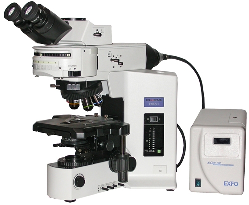
- In a fluorescence microscope (like the one in the top picture), we stain the specimen with a fluorescent dye, and bombard it with light of a particular wavelength (usually from a mercury lamp).
- A fluorescence microscope is much the same as a conventional light microscope with added features to enhance its capabilities.
- Fluorescence microscopes can observe cells and structures either emitting auto fluorescent or stained by the fluorochromes.
- Fluorescence microscopes have evolved together with the development of fluorescent dyes around the early 20th century.
- Fluorescence microscopes and DIC (alternativeial interference contrast) are other types of compound microscopes.
- The fluorescence microscope is used to examine structures that bind special fluorescent dyes.
- Fluorescence microscopes are especially useful in clinical microbiology.
History of Fluorescence microscopes
- In 1978 first theoretical ideas have been developed to break this barrier by using a 4Pi microscope as a confocal laser scanning fluorescence microscope where the light is focused ideally from all sides to a common focus which is used to scan the object by ‘point-by-point’ excitation combined with ‘point-by-point’ detection.[8] However, the first experimental demonstration of the 4pi microscope took place in 1994.[9] 4Pi microscopy maximizes the amount of available focusing directions by using two opposing objective lenses or two-photon excitation microscopy using redshifted light and multi-photon excitation.
- In the 1930s, florescent staining was applied to investigating and observing the morphology of bacteria, mildew, cells, and fiber.
- In the 1940s, the fluorescent staining protein technology was invented and applied extensively to the routine clinical immuno-fluorescence staining, which can examine and locate virus, bacteria, mildew, protozoan, parasite, antigen, and antibody in animals and humans.
- In the 1990s, the laser scanning confocal fluorescence microscopy was developed.