Contents
- 1 Overview of Simple microscopes
- 2 A Five-Year Old Inventor?
- 3 But I only have $50-100 to spend; does that mean no microscopy?
- 4 Can I check lens quality?
- 5 How does a simple microscope work?
- 6 I need compound scopes for my class, and I see a lot of features advertised; which ones are worthwhile?
- 7 I want to equip my classroom with “real” microscopes; what will that cost?
- 8 Microscope History – Who Invented the Microscope?
- 9 What are the mechanical parts?
- 10 What are the optical parts?
- 11 What features should I look for?
- 12 What is a simple microscope used for?
- 13 What is a simple microscope?
- 14 What is Microscopy?
- 15 What is the difference between compound microscope and simple microscope?
- 16 What is the principle of a simple microscope?
- 17 What kind of microscope should I buy?
- 18 What type should I buy?
- 19 Where do I find these microscopes?
- 20 History of Simple microscopes
Overview of Simple microscopes
- Simple microscopes are gaining popularity again with 2D/3D folded and printed versions that are easily constructed, providing an inexpensive instrument that can be used in harsh environments which include disease identification in disaster or emergency relief situations.
- A simple microscope is essentially a magnifying glass made of a single convex lens with a short focal length, which magnifies the object through angular magnification, thus producing an erect virtual image of the object near the lens.
- The simple microscope is a converging optical system—often a simple lens—that makes it possible to reduce the observation distance and thus to increase the apparent size of the object.
- A simple microscope is a useful tool in microbiology, as it can be used to view microscopic organisms and other biological specimens which include algae, fungi, protists, and Hydra.
- A simple microscope is a rudimentary magnification device that is capable of visibly enlarging small objects, so they can be viewed and studied in better detail.
- Simple microscopes relied on natural light around the object or a direct form of illumination passing right through it if it was translucent.
- A simple microscope is simply a large magnifying glass with a shorter focal length that has a convex mirror with a small focal area.
- Simple microscopes consist of a single lens mounted in a holder that is either fixed, or can be adjusted for focusing.
- A simple microscope is a microscope that uses only one lens for magnification, and is the original light microscope.
- Simple microscopes may contain a reticle (used to verify the size of objects).
A Five-Year Old Inventor?
The compound microscope was created and developed between the 16th and 17th centuries. It is generally accepted that Zaccharias Janssen (1585-1632), an eyeglass maker from the Netherlands, is the inventor, but he was five to ten years old at the time of invention (around 1590 to 1595). One theory is that he helped his father in their workshop and the credit should go to them both. Their microscope consisted of a handheld central cylinder with moveable eyepiece and objective tubes (Figure 1). The bi-convex eyepiece and plano-convex objective lenses produced a magnification of between three and nine times.
But I only have $50-100 to spend; does that mean no microscopy?
Not at all! Many manufacturers offer a design that looks like a pocket flashlight. They usually magnify 30x, with two AA batteries providing illumination, and they sell for about $10. You’ll find them in electronics and “nature” stores, and many catalogs; quality varies, so it’s wise to compare. They can be good enough to support extensive curriculum. Buy as many as you can afford; some local dealers may be willing to discount a bulk purchase for school use.
Can I check lens quality?
YES. You can tell a lot without test equipment. The rectangular engraved crosshatching around the portrait heads on U. S. currency is a useful specimen for a crude check of lens quality.
How does a simple microscope work?
Simple microscopes make use of a biconvex lens to magnify the image of a specimen. Nowadays, these lenses often consist of two glass elements with color correction abilities. The closer the object is to the lens, the larger the magnified image becomes.
I need compound scopes for my class, and I see a lot of features advertised; which ones are worthwhile?
“Magnifies 600-1200 times!” NO. When you see this claim in an advertisement, it’s good reason to read no further. The wavelength(s) of visible light and the optical properties of glass lenses used in air (rather than the “immersion oil” used with research microscopes) limit the useful magnification of a compound school microscope to 400x; more is “empty magnification”. Magnification can be calculated by multiplying the power of the eyepiece lens by that of the objective lens. For example, using a 10x eyepiece and a 40x objective gives 400x. Higher magnifications are achieved in “toy” microscopes by using an eyepiece of excessive power, which in turn makes the field of view very narrow, while emphasizing all the aberrations of the image produced by the objective lens. It’s like enlarging a snapshot from a cheap camera to poster size; it’s bigger, but there’s no more detail. Most school microscopy needs 10-100x (bacteria require 400x). True 1000x imaging requires a 4th objective (100x) in the turret, a multi-lens focusable condenser, plus the use of immersion oil. It should only be considered for advanced high school classes.
I want to equip my classroom with “real” microscopes; what will that cost?
Approximately $1000 (don’t despair; see below). That will get you at least 10 good quality scopes in the $80-150 price range. You can get scopes at that cost that will be durable and easy to use, with lenses that will deliver a sharp, bright image. In general, more expensive models will provide similar images but more convenience, and less expensive ones will have disappointing performance.
Microscope History – Who Invented the Microscope?
During the 1st century AD (year 100), glass had been invented and the Romans were looking through the glass and testing it. They experimented with different shapes of clear glass and one of their samples was thick in the middle and thin on the edges. They discovered that if you held one of these “lenses” over an object, the object would look larger.
What are the mechanical parts?
Mechanical parts pertain to the parts of the microscope that support the optional parts. They help in the adjustment so as to accurately magnify the object.
What are the optical parts?
They are the parts of the microscope that involved passing the light through the specimen and magnify its size.
What features should I look for?
Both types should have metal bodies and metal rack-and-pinion focus, for durability and easy, precise focusing. That immediately eliminates the plastic “toy” scopes. Although a metal body is no guarantee of lens quality, metal focus gearing is more precise than twistable or plastic designs. Both types should have glass rather than plastic lenses and be able to focus on both thin specimens (slides) and the surface of larger objects at least an inch thick. Compound scopes should have a 3-lens turret and a substage diaphragm or series of “field stops” to control brightness. There are some good single-objective compound scopes available, but the three lens design is much more versatile; a student can locate a subject at low power and immediately switch to higher magnifications of the same area.
What is a simple microscope used for?
While simple microscopes are, well, simple imaging devices, these microscopes still have a variety of uses and applications, from scientific studies like biology, to more practical activities such as jewelry and watchmaking, and in looking at books, cloths, stamps, and engravings.
What is a simple microscope?
A simple microscope is essentially a magnifying glass made of a single convex lens with a short focal length, which magnifies the object through angular magnification, thus producing an erect virtual image of the object near the lens.
What is Microscopy?
Microscopy is the art of using microscopes to find, examine, and discover things that can’t be seen with the naked eye. Our first use of microscopes involved lenses for physical magnification, but now we have access to complex microscopes that allow us to see well beyond physical lenses. These different types of microscopes fall into three common categories- optical microscopy, electron microscopy, and scanning probe, but a fourth, x-ray, is also in the mix. Optical microscopes, also called light microscopes, involve light passing through lenses and the specimen, providing magnification. They include simple microscopes and compound microscopes. You likely learned about those fundamentals (i. e. , a field of view, light sources, eyepieces, and sample preparation) in high school. Electron microscopes use the same principle, but shine electron beams through the specimen, allowing for sharper images of even smaller things. They fall into two categories, transmission electron microscopy, and scanning electron microscopy. They go beyond visible light to produce 3D images in some cases. Scanning probe microscopy uses a probe to scan the surface of a specimen for great detail. Image analysis and microscopy techniques have moved beyond even these three to produce high-quality images of things we never thought we’d see.
What is the difference between compound microscope and simple microscope?
As the name suggests, a simple microscope uses a single lens for magnification while a compound microscope uses various lenses to further magnify the object.
What is the principle of a simple microscope?
If you place a tiny object within the focus of the simple microscope, a magnified image of the object is formed making it easier for the naked eye peeping through the lens to see the image.
What kind of microscope should I buy?
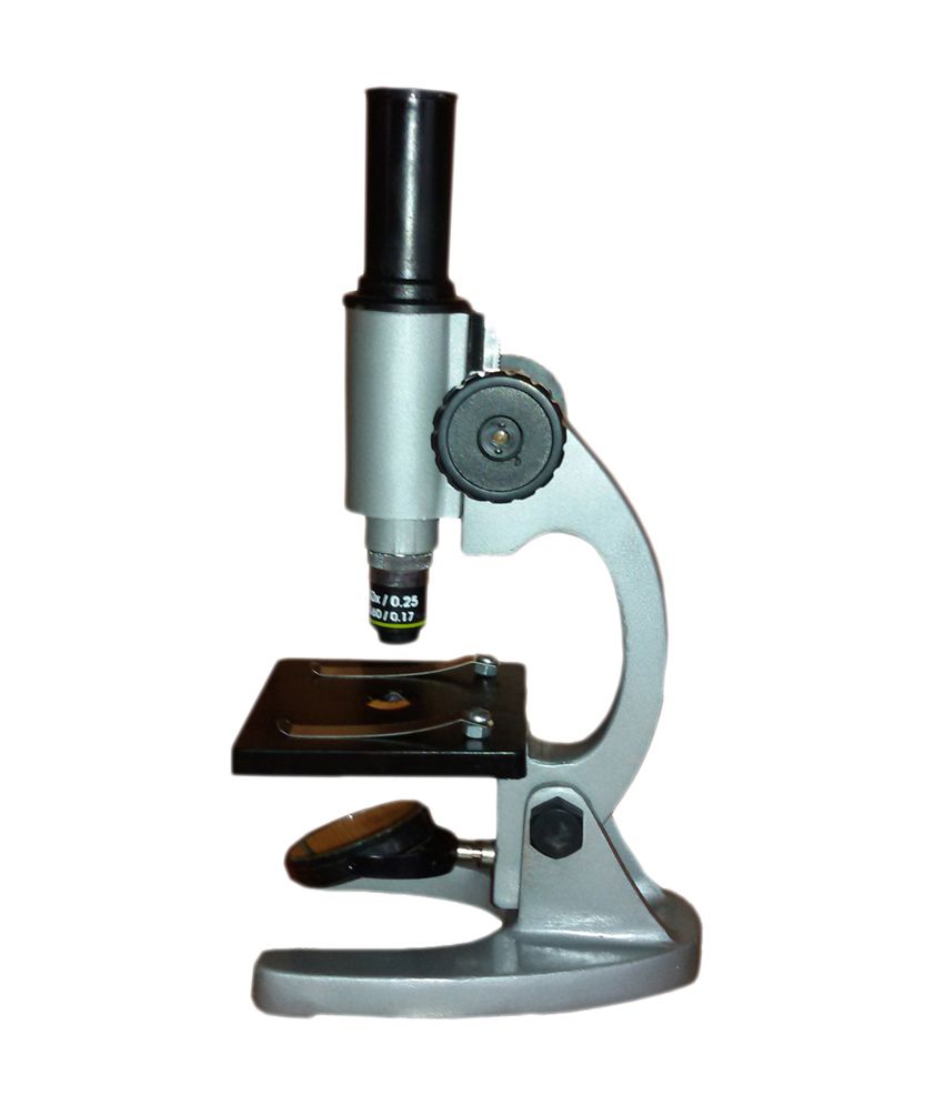
The first choice is between “simple” and “compound” microscopes. A “simple” microscope (Leeuwenhoek used one) has just one lens and a “compound” scope has both an objective and an eyepiece. Don’t buy a “simple” design! The working distances between eye and lens and lens and specimen are so small that they are very difficult to use. And a single powerful lens has so much aberration that the student who manages to get an image will be disappointed by its quality. Unfortunately, there are quite a few models offered in school supply catalogs.
What type should I buy?
Two types, actually, in roughly equal numbers for middle school. Inspection/dissection scopes are used to look at surface details of large, opaque specimens at relatively low (20-30x) power. Illumination is usually from above, and the image is erect, as in the “real world”. Compound microscopes are usually used with transmitted light to look through transparent specimens; the useful school magnification range is 10-400x. The image is inverted. It takes a bit of practice to follow a moving subject when it’s upside-down.
Where do I find these microscopes?
Major scientific supply catalogs and some of the school supply houses will have them; this web page has a dealer contact list. A microscopes-only dealer may provide both a presale quality-control check and in-house service. Shop carefully; prices may vary by 50% or more.
History of Simple microscopes
- In 1665, Robert Hooke used an improved compound microscope to observe cells.
- In the 1660s, another Dutchman, Antonie van Leeuwenhoek (1632-1723) made microscopes by grinding his own lenses.
- In the 1850s, John Leonard Riddell, Professor of Chemistry at Tulane University, invented the first practical binocular microscope while carrying out one of the earliest and most extensive American microscopic investigations of cholera.

